Sinusitis, a condition that inflames the tissues around the sinuses, stands as one of the most common health care complaints worldwide. Its persistent prevalence makes it a significant focus for health care providers, particularly family practitioners, who often diagnose the condition based on patient history and physical examinations.
In many instances, patients respond to medical management, and no further imaging becomes necessary. However, certain situations call for more advanced diagnostics like X-rays, offering detailed insight into the patient’s paranasal sinus pathology. This comprehensive guide will delve into the techniques used for Paranasal Sinuses (PNS) X-Rays, highlighting the importance of Water’s, Caldwell, lateral, and basal views.
Overview of Sinusitis and the Need for X-Rays
Sinusitis can be acute or chronic, with each type presenting varying degrees of discomfort and symptoms. These symptoms often include nasal inflammation, sinus pressure, headache, and even fever. The ability to accurately diagnose and treat sinusitis relies heavily on understanding the patient’s symptoms and underlying causes. Family practitioners are adept at diagnosing and treating sinusitis based on symptoms and physical examinations. However, persistent or severe cases may necessitate the use of imaging diagnostics.
Plain radiography or X-rays are often employed to diagnose paranasal sinus pathology. X-ray imaging uses a small amount of radiation to produce images of the inside of the body, allowing physicians to examine the structure and function of the sinuses in detail. This is particularly beneficial in identifying any abnormalities or pathologies.
Paranasal Sinus X-Ray Techniques
To capture the most detailed and informative images of the sinuses, radiologists utilize various viewing techniques. Four of the most common include Water’s, Caldwell, lateral, and basal views.
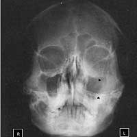
Water’s View (Occipito-mental)
Considered the primary view for observing the maxillary antra and the anterior ethmoid sinuses, the Water’s view is essential for a comprehensive X-ray examination of the paranasal sinuses. Besides the maxillary antra and the anterior ethmoid sinuses, the Water’s view also well represents the orbits, nasal cavities, and zygomatic arches.
Importance and Utility of the Water’s View
The Water’s view is particularly beneficial due to its wide coverage of sinus areas. The maxillary sinuses, situated in the cheek area on each side of the nose, are the largest sinuses. Any infections or blockages here may lead to sinusitis symptoms such as facial pain and pressure. By providing a clear view of the maxillary antra, the Water’s view aids in diagnosing these conditions.
In addition, the Water’s view allows physicians to examine the anterior ethmoid sinuses, which are located between the eyes. These sinuses are often affected by colds and allergies, leading to sinusitis symptoms like headaches between the eyes.
The Procedure for the Water’s View
For the Water’s view, the patient is usually asked to lean against the X-ray machine, their chin extended forward. This position helps to bring the facial bones and sinuses into a clear view for the X-ray. The resulting image provides a detailed representation of the maxillary antra, the anterior ethmoid sinuses, the orbits, nasal cavities, and zygomatic arches.
Caldwell View (Occipito-frontal)
The Caldwell view highlights the posterior ethmoid cells and frontal sinuses. The frontal sinuses, located in the forehead, can cause a great deal of discomfort when infected or inflamed. The Caldwell view offers an in-depth look into these sinuses, facilitating accurate diagnosis and treatment.
Importance and Utility of the Caldwell View
The Caldwell view offers a clear image of the posterior ethmoid cells and the frontal sinuses. Frontal sinuses, positioned behind the forehead, can cause discomfort or headaches across the forehead when inflamed or infected. Therefore, by providing a detailed image of these sinuses, the Caldwell view plays a crucial role in diagnosing such conditions.
The posterior ethmoid sinuses, located near the back of the nose, are also well represented in this view. They can be involved in more serious sinusitis symptoms such as pain in the top of the head or around the temples. By offering a clear view of the posterior ethmoid cells, this view aids in diagnosing any inflammation or infection in these areas.
The Procedure for the Caldwell View
In the Caldwell view procedure, the patient is usually positioned facing the X-ray machine with their forehead and nose touching the device. The central X-ray beam is directed through the forehead, creating a clear image of the posterior ethmoid cells and the frontal sinuses.
Lateral View
The lateral view technique is an X-ray taken from the side of the patient’s face, which captures the sinuses in a unique orientation. This view is specifically employed to display any air-fluid level in the sphenoid sinus. Besides the sphenoid sinus, the sella turcica and the nasopharynx are also well-seen in this view.
Importance and Utility of the Lateral View
The lateral view can provide a comprehensive picture of the sinuses, particularly the sphenoid sinuses. Located deep within the skull, the sphenoid sinuses can be difficult to diagnose and treat. Sinusitis affecting these sinuses may cause symptoms like a deep headache or pain at the back of the head. The lateral view gives an effective representation of these sinuses, aiding in accurate diagnosis and treatment.
In addition, the lateral view can help detect abnormalities in the sella turcica, a saddle-shaped area in the sphenoid bone. The nasopharynx, the area behind the nose and above the back of the throat, can also be examined using this view.
The Procedure for the Lateral View
In the lateral view procedure, the patient’s head is turned to the side, and the X-ray beam is directed from one side of the head to the other. This angle allows the X-ray to provide a clear image of the sphenoid sinus, sella turcica, and nasopharynx.
Basal View
The basal view, also known as the submentovertex view or Hirtz compass view, provides a comparison of each sphenoid sinus and also visualizes the ethmoid sinuses and nasal cavities.
Importance and Utility of the Basal View
The basal view allows for a comparison of both sphenoid sinuses, helping to identify any abnormalities or asymmetries between them. This can be crucial for the diagnosis of conditions affecting these sinuses. This view also offers a comprehensive view of the ethmoid sinuses and the nasal cavities, facilitating the identification of any abnormalities in these areas.
The Procedure for the Basal View
In the basal view procedure, the patient’s head is tilted back, and the chin is pointed upward. The X-ray beam is directed from the bottom (base) of the skull toward the top, offering a view of each sphenoid sinus, the ethmoid sinuses, and the nasal cavities.
Final Thoughts
Given the vast and diverse symptoms of sinusitis, having a comprehensive and effective method for diagnosis is critical. With techniques such as Water’s, Caldwell, lateral, and basal views, Paranasal Sinuses X-Rays offer valuable insight into the condition of a patient’s sinuses, helping physicians to diagnose and treat sinusitis more effectively.
While the Water’s view offers clear images of the maxillary antra and anterior ethmoid sinuses, the Caldwell view puts the spotlight on the posterior ethmoid cells and frontal sinuses. On the other hand, the lateral view allows for a unique orientation to showcase the sphenoid sinus, sella turcica, and nasopharynx, while the basal view provides comparative images of each sphenoid sinus and visualizes the ethmoid sinuses and nasal cavities.
Each of these views contributes to forming a comprehensive understanding of the patient’s sinus condition. Therefore, it’s crucial that medical professionals understand the specifics of these techniques, their importance, and the procedures involved.
As with any medical procedure, the comfort and safety of the patient are paramount. When conducting PNS X-rays, it’s important to ensure that the patient is comfortable and understands what the procedure entails. Also, because X-rays use radiation, it’s critical that the procedure be conducted with the utmost care to ensure minimal exposure.
In conclusion, PNS X-rays play a vital role in diagnosing sinusitis and other related conditions. Understanding the different views and their importance is key to ensuring accurate diagnoses and effective treatments. While technology continues to advance and provide new and improved diagnostic tools, PNS X-rays remain a valuable, time-tested tool in the field of medical diagnostics. Their role in detecting and diagnosing sinus conditions is indisputable, making them a critical component in the management and treatment of sinusitis.
No matter the advancements in medicine, the aim remains the same – to ensure the highest standard of care for patients. Therefore, being equipped with the knowledge and understanding of diagnostic tools like PNS X-rays is essential for every healthcare provider.
Enhancing Diagnostic Accuracy with PNS X-Rays
The accurate interpretation of PNS X-rays relies on the expertise of the radiologist and the clarity of the images produced. Each view offers unique angles and perspectives on the sinuses, giving a well-rounded picture of the patient’s condition. However, no single view can provide all the information needed. The combination of these various views, each illuminating different areas, contributes to the creation of a holistic understanding of the patient’s sinus condition.
Understanding and interpreting these X-ray views correctly is crucial in the diagnostic process. Misinterpretation can lead to incorrect or missed diagnoses, which can delay appropriate treatment. Thus, thorough training and constant practice are necessary for any healthcare provider dealing with these types of X-rays. It is also essential to stay updated on any new techniques or advances in the field that might improve the effectiveness and accuracy of these diagnostic tools.
Patient Communication and Consent
Before conducting PNS X-rays, it’s crucial to ensure the patient fully understands the process and consents to it. The healthcare provider should explain why the X-ray is necessary, what it involves, and any risks associated with it. Patient anxiety can be significantly reduced when they are well-informed about the procedures they’re undergoing.
Additionally, it’s important to consider the patient’s comfort during the procedure. Positioning for these X-rays can sometimes be uncomfortable, but careful handling and reassurances can help make the process as easy as possible for the patient.
The Future of Sinus Imaging
While PNS X-rays continue to be a common diagnostic tool, technological advancements have introduced other imaging techniques, such as computed tomography (CT) and magnetic resonance imaging (MRI). These methods provide more detailed images and can sometimes offer more information about the soft tissues around the sinuses. They are often used in more complicated cases or when more detailed images are needed to guide surgical procedures.
Despite these advancements, the simplicity, accessibility, and effectiveness of PNS X-rays make them a staple in sinus diagnostics. They are an affordable and quick way to gain valuable insight into the patient’s condition and form an initial understanding that can guide further diagnostics and treatment.
Ultimately, the goal of any diagnostic tool is to aid in the accurate diagnosis and effective treatment of conditions. In the case of sinusitis and other sinus-related conditions, PNS X-rays are an invaluable tool in achieving this goal. The four primary views—Water’s, Caldwell, lateral, and basal—are instrumental in identifying and assessing sinus pathology, guiding treatment decisions, and improving patient outcomes.
Healthcare providers, radiologists, and all those involved in the diagnostic process must be adept in understanding and interpreting these views, ensuring that they continue to provide the vital link between symptoms and treatment that they have for many years. With their help, patients around the world can continue to receive accurate diagnoses and effective treatments for their sinus conditions.
pathology pns in x.ray view
Pns om view